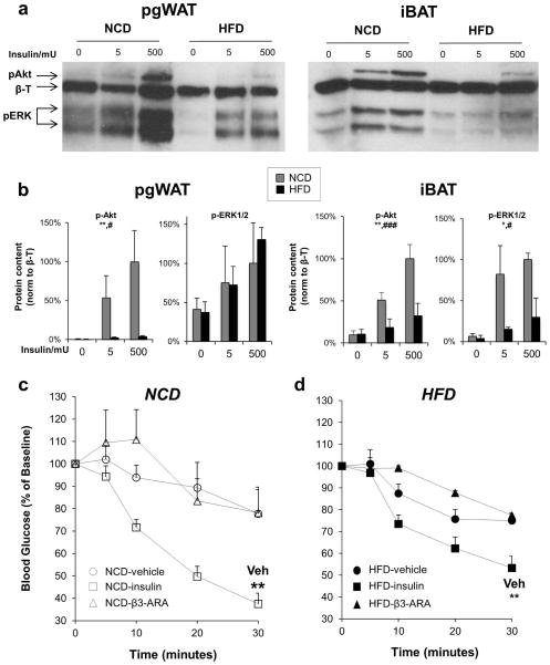
Figure 2. High fat diet reduces insulin signaling in pgWAT and iBAT.
(a) Representative immunoblots of phospho-Akt (S473) and phospho-ERK in pgWAT and iBAT from mice on NCD or HFD for 18 weeks treated via IVC injection with vehicle, 5 mU insulin, or 500 mU insulin for 5 minutes before tissue harvest. β-Tubulin (β-T) was used as a loading control. Data shown are representative of three independent experiments. (b) Densitometry of p-Akt (n=4) and p-ERK (n=2). Two-way ANOVA *P < 0.05, **P < 0.01 for comparing NCD and HFD, and #P < 0.05, ###P < 0.001, for comparing insulin doses. Blood glucose levels were determined after injection of vehicle (circles), 0.75 mU/kg insulin (squares), 0.1 μg/g body weight of the β3-adrinergic agonist mirabegron (triangles) in mice fed (c) NCD (white symbols) or (d) HFD (black symbols). n=5–6 per treatment. **P < 0.01, compared with vehicle.

