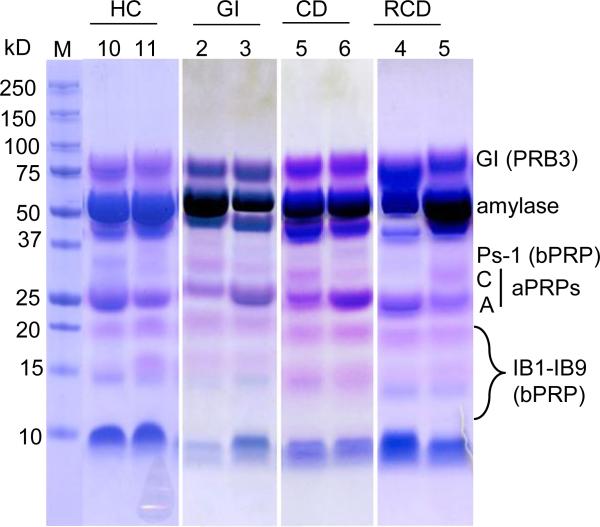
Figure 4.
SDS-PAGE (12% gel) of PS from two patients of each group. Aliquots of 35 μL PS were loaded for each subject. The gel was stained with Coomassie Blue R-250 using a modified destaining method [36]. The PRPs stain pink or violet while other proteins stain blue.

