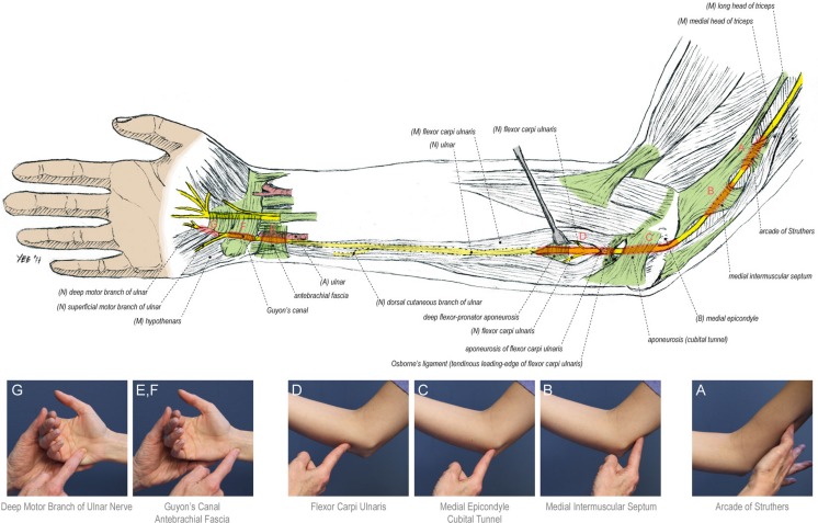
Fig. 1.
Ulnar nerve anatomy, course, and sources of compression in the upper extremity. Clinical photographs indicate the surface landmarks for the sites of ulnar nerve compression tested in this study, which include the following: (1) the arcade of Struthers (a), a transverse attachment of the deep fascia of the distal triceps to the medial intermuscular septum, located approximately 8 cm proximal to the medial epicondyle (2.22–24); (2) the medial intermuscular septum (b); (3) the bony retrocondylar groove and the overlying cubital tunnel retinaculum spanning the olecranon and medial epicondyle (c); (4) Osborne’s band (d), a thickened confluence of fascia between the two heads of the flexor carpi ulnaris muscle (5–6); (5) the volar antebrachial fascia just proximal to the wrist crease (e, f); and (6) the tendinous leading edge of the hypothenar musculature overlying the deep motor branch of the ulnar nerve [8] (g)

