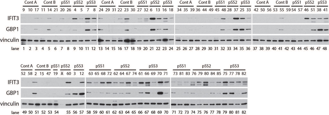
Figure 1. Distinct patterns of IFN activity are evident in lysates made from LSG biopsies from SS participants.
Protein lysates made from control (n=29) or SS (n=53) LSG biopsies were probed for IFN activity by Western blotting. A marker of type I IFN (IFIT3) and type II IFN (GBP1) is included. Vinculin was analyzed as a loading control.

