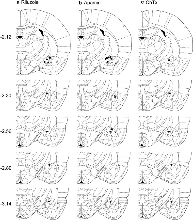
Fig. 4.

Location of microdialysis probes for stereotaxic drug application. Symbols show the positions of the tips of microdialysis probes for drug application into amygdala regions. CeA, filled circles; BLA, open circles. a Positions of microdialysis probes for stereotaxic application of riluzole into the CeA. b Positions of microdialysis probes for stereotaxic application of apamin into CeA or BLA; gray symbols indicate sites of apamin injection that increased vocalizations. c Positions of microdialysis probes for stereotaxic application of charybdotoxin (ChTx) into CeA or BLA. Diagrams show coronal brain slices. Numbers indicate distance from the bregma
