
Fig. 1.
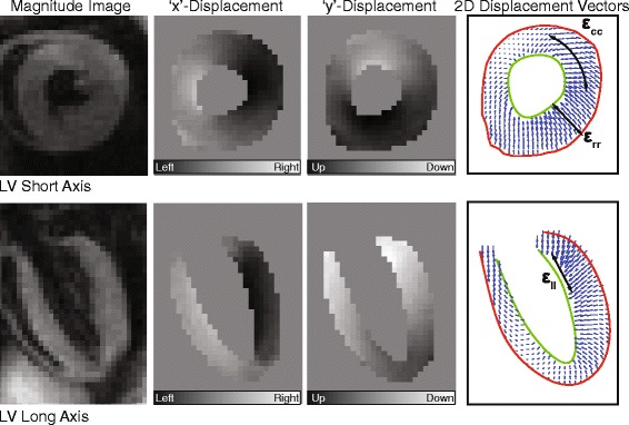
Representative DENSE CMR images of a mouse left ventricle (LV) in the short (top) and long axis (bottom) orientations at peak systole. The far right panel demonstrates the corresponding vector displacement field that can be derived from the phase displacement images (middle panels), along with the corresponding decomposition into radial (εrr), circumferential (εcc), and longitudinal (εll) strains