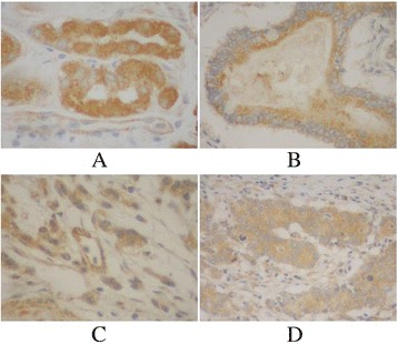
Fig. 5.

Immunohistochemical staining for Hoxd10 in gastric cancer lesions and noncancerous tissues, magnification 400×. a Hoxd10 was highly expressed in noncancerous tissues. b Hoxd10 was highly expressed in tubular adenocarcinoma. c Hoxd10 was highly expressed in poorly differentiated adenocarcinoma. d Hoxd10 was highly expressed in poorly differentiated adenocarcinoma.
