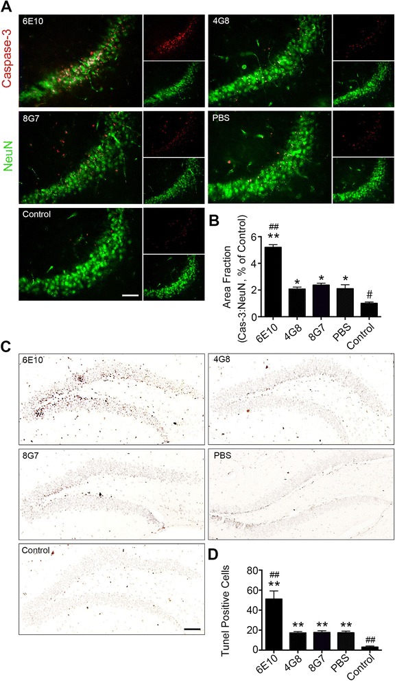
Fig. 3.

Disaggregation of preformed Aβ fibrils by an N-terminal antibody increases the neurotoxicity of Aβ in vivo. a Neuronal apoptosis in the CA3 region was visualized using NeuN and activated caspase-3 co-staining (scale bar = 50 μm). b Quantification of apoptosis by activated caspase-3 in the CA3 region. c Neuronal apoptosis in the dendrite gyrus was visualized using TUNEL staining (scale bar = 200 μm). d Quantification of apoptotic cells in the dendrite gyrus that were stained by TUNEL (n = 5 per group, mean ± SEM, One-way Anova,*P < 0.05 and **P < 0.01 vs control, #P < 0.05 and ##P < 0.01 vs PBS)
