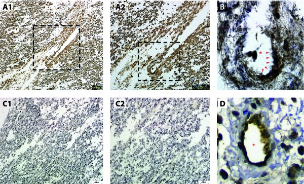
Figure 1. A1: Low power (10 ×) section of autopsied human spinal cord tissue from active lesion in a patient with neuromyelitis optica demonstrating diffuse perivascular c4d complement deposition. A2: Boxed area is magnified in A2. Higher power section (20 ×) of boxed area from A1 showing perivascular C4d staining. B: High power section (40 ×) of boxed area from A2 showing intense perivascular C4d staining (arrows). Asterisk identifies the blood vessel lumen. C1: Low power (10 ×) section of spinal cord without primary antibody against C4d showing counterstain only. C2: Higher power section (20 ×) from C1 confirming no C4d staining. D: Image from transplant rejected heart tissue showing classical perivascular c4d staining pattern. Asterisk identifies the blood vessel lumen.

