
Figure 2.
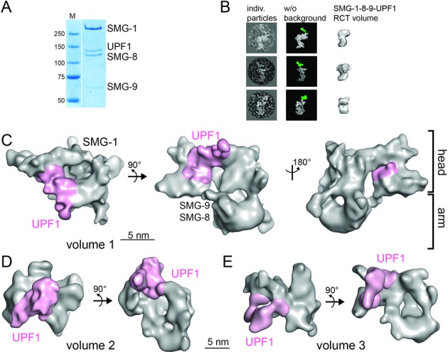
Cryo-EM of the SMG-1–8–9-UPF1 complex. (A) Coomassie-stained SDS-PAGE gel of purified SMG-1–8–9-UPF1. (B) Localization of UPF1 in the SMG-1–8–9-UPF1 complex using a polyclonal antibody against the UPF1 CH domain. Individual particles showing the anti-UPF1 antibody bound to the complex (left) are depicted next to the same particles after background removal (middle). Density corresponding to the antibody is colored in green. The RCT reconstruction of SMG-1–8–9-UPF1 is shown in a similar orientation (right). (C) Cryo-EM structure of SMG-1–8–9-UPF1 complex volume 1. SMG-8, SMG-9 and UPF1 are positioned based on results from antibody-labeling experiments ((23,31), this study). Density corresponding to SMG-1–8–9 is depicted in gray and UPF1 in magenta. (D and E) Cryo-EM structures of SMG-1–8–9-UPF1 volume 2 (D) and volume 3 (E) with UPF1 density colored in magenta.