
Figure 7.
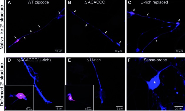
Validation of structural requirement of the zipcode RNA for axonal localization. Panels (A)–(C) showed representative images of DRG neurons expressing GFPmyrzipcodewild-type, GFPmyrzipcodeΔACACCC, and GFPmyrzipcodeU-rich replaced constructs, respectively. GFP mRNA (red) and protein (blue) signals were observed in both the axons and growth cones (arrows) of DRG neurons transfected with reporter constructs containing the native-like secondary structure. However, those with the disrupted secondary structure [D; GFPmyrzipcodeΔ(ACACCC/U-rich), E; GFPmyrzipcodeΔU-rich] showed reporter mRNA in the cell body only (asterisks represented cell body in insets). Sense cRNA riboprobe for GFP showed no signal in exposure-matched images of GFPmyrzipcodewild-type transfected DRGs (F).