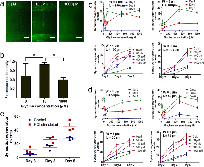
Figure 3. Induction of functional synaptic communication by glycinergic factors and potassium chloride (KCl).
(a) Immunostaining image of phosphorylated extracellular-related kinase (pERK) in retinal cells treated with glycine at concentrations of 0, 10 and 1000 μM. Scale bar, 50 μm. (b) Histogram of fluorescence intensity of pERK and glycine concentrations at 0, 10 and 1000 μM. *p < 0.05. (c–d) Effect of microchannel width (4 μm and 3 μm) with channel length of 100 μm (c) and 50 μm (d) on retinal synaptic regeneration at glycine concentrations of 0, 50, 100, 500 and 1000 μM. W: channel width. L: Channel length. (e) Dynamics of chemically induced retina synaptic regeneration via the stimulation of KCl, compared to control sample without KCl treatment on days 3, 5 and 8. The data in b and c represents the mean ± s.e.m. with n = 3.

