
Figure 2. Single-projection phase contrast and attenuation contrast images of a mouse pup specimen suspended in a water-filled chamber.
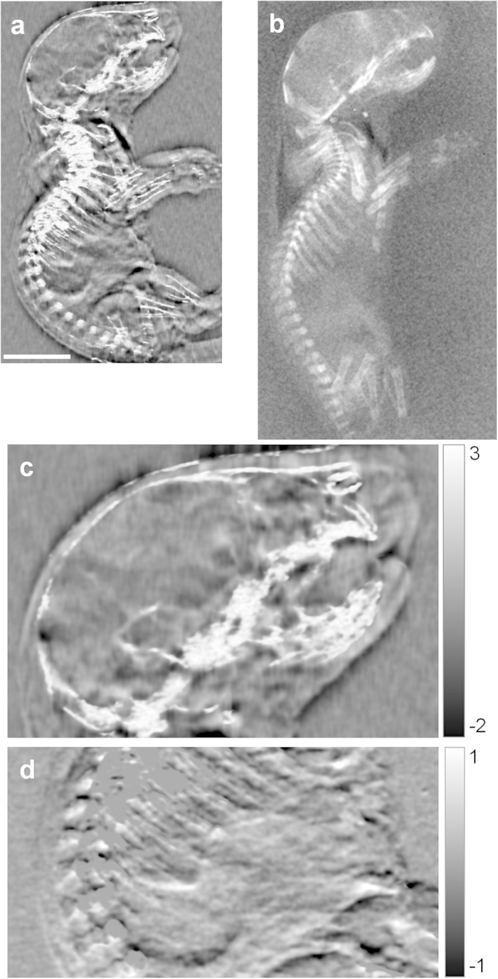
(a) Phase contrast image from the PFI at 1.08 mGray entrance surface radiation dose. The phase contrast image is a fused image of the phase shift of the wavefront and the linear intensity attenuation (see Methods section for description). The scale bar is 5.0 mm. (b) Attenuation contrast image from a digital flat panel detector at 3.08 mGray dose. (c) Magnified view of the head in the fused phase contrast image. Grayscale unit is radians. (d) Differential phase data of the abdomen area. Grayscale unit is radians. The highly scattering bones are removed by thresholding the scatter signal.