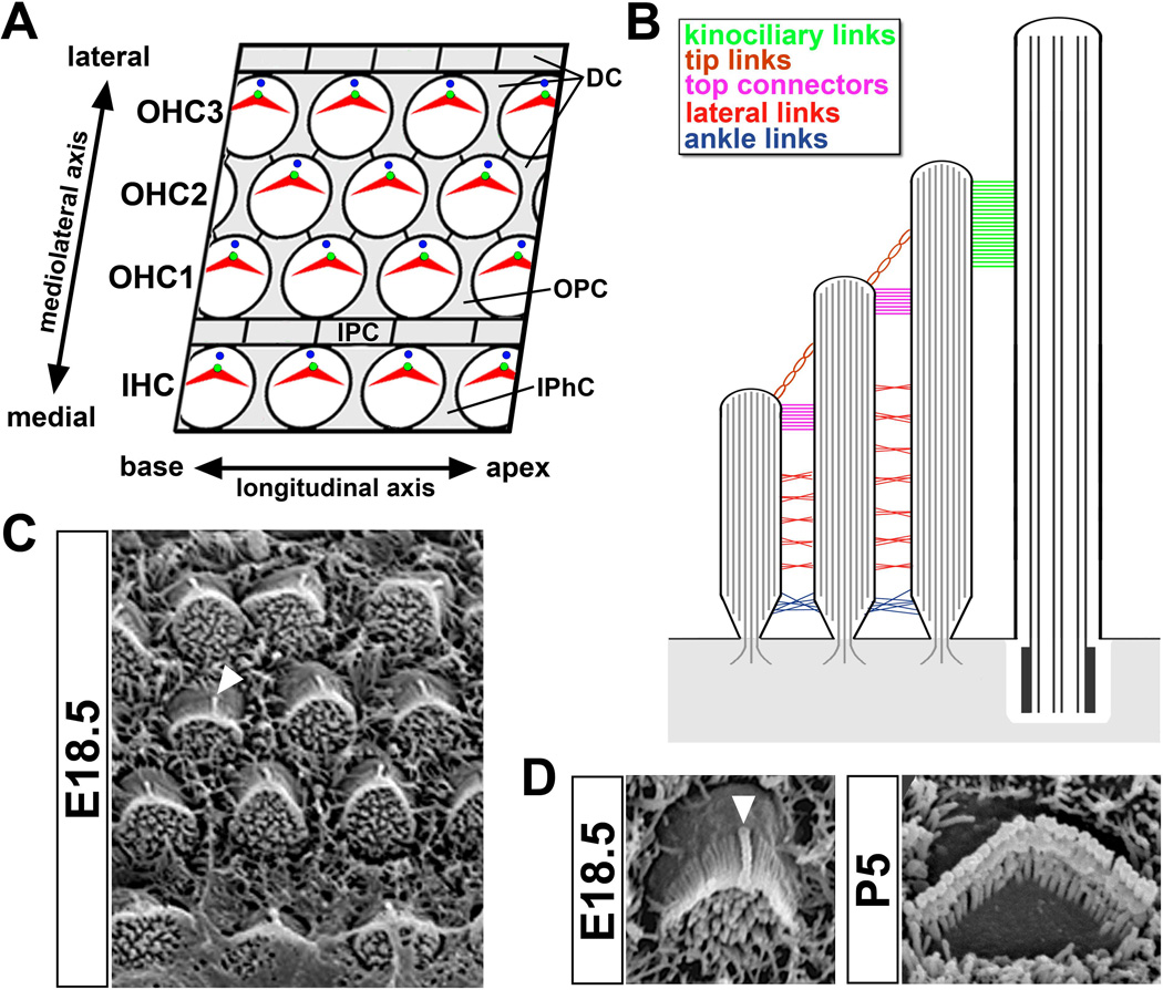
Figure 2. Organization of the organ of Corti and hair bundle.
(A) En face diagram of the mammalian organ of Corti showing the mosaic pattern of hair cells (shaded white) and supporting cells (shaded gray). The mediolateral and longitudinal axes are indicated. Abbreviations indicate distinct cell types: IHC, inner hair cell row; OHC1–3, outer hair cell row; DC, Deiters’ cell; OPC; outer pillar cell; IPC, inner pillar cell; IPhC, inner phalangeal cell. (B) Cross-sectional diagram of the hair bundle depicting five distinct populations of stereociliary links colored according to the accompanying key. Stereocilia rootlets are anchored in the cuticular plate (shaded gray). The kinocilium and underlying basal body are on the right. (C) En face scanning electron micrograph of the mouse organ of Corti at E18.5. White triangle indicates the kinocilium of an individual hair cell. (D) Left, an E18.5 hair bundle showing the kinocilium (white triangle) lying at the vertex of the hair bundle. Right, a P5 bundle showing the mature V-shape and staircase arrangement of the three stereocilia rows in an outer hair cell. Note that the kinocilium has been resorbed.

