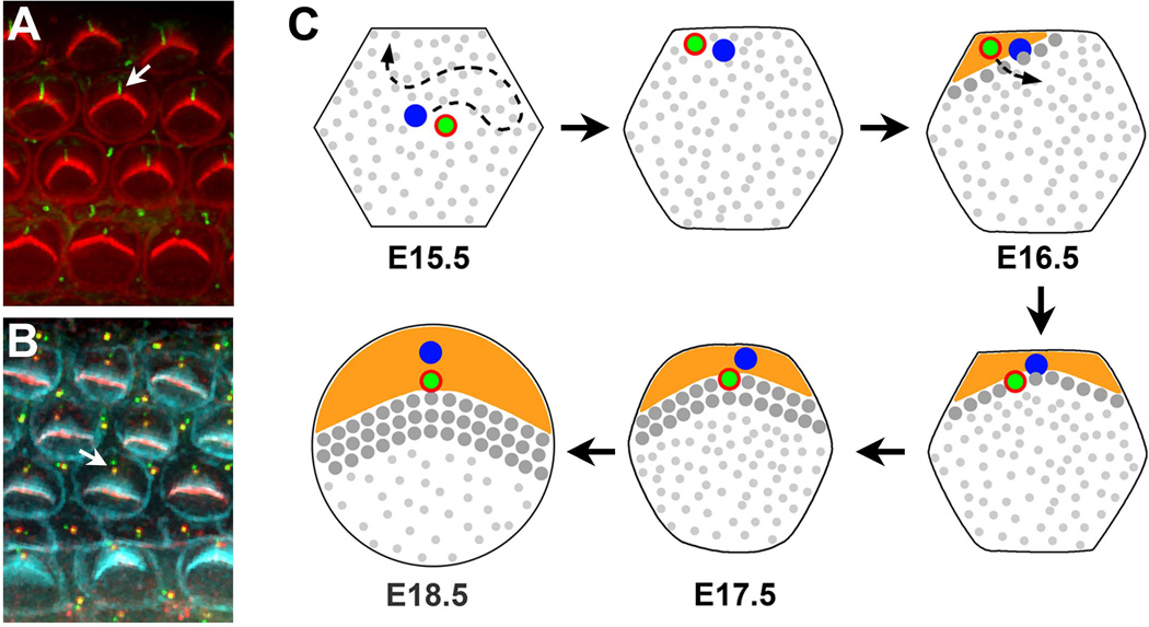
Figure 3. Development of planar cell polarity in the organ of Corti.
(A) En face view of the organ of Corti. Arrow indicates the kinocilium at the hair bundle vertex. Acetylated tubulin, green; actin, red. (B) Planar-polarized arrangement of the basal body and the associated daughter centriole along the medial-lateral axis (arrow). The basal body is labeled by phospho-β-catenin staining (red) and centrioles are labeled by Centrin2-GFP (green). F-actin, cyan. (C) Schematic diagram of kinocilium/basal body movements and early apical morphogenesis in the mouse. At E15.5, the centrally placed kinocilium/basal body (green dot with red annulus) migrates to the cell periphery where it closely associates with the cortex. A bare zone (orange) devoid of microvilli (small gray dots) subsequently develops in the vicinity of the kinocilium. As the bare zone expands, the kinocilium/basal body relocalizes to a more central position where it aligns along the medial-lateral axis with the daughter centriole. Here, the nascent hair bundle forms with the kinocilium positioned at its vertex (large gray dots), abutting the expression domain of bare zone proteins. Daughter centrioles, blue dots.

