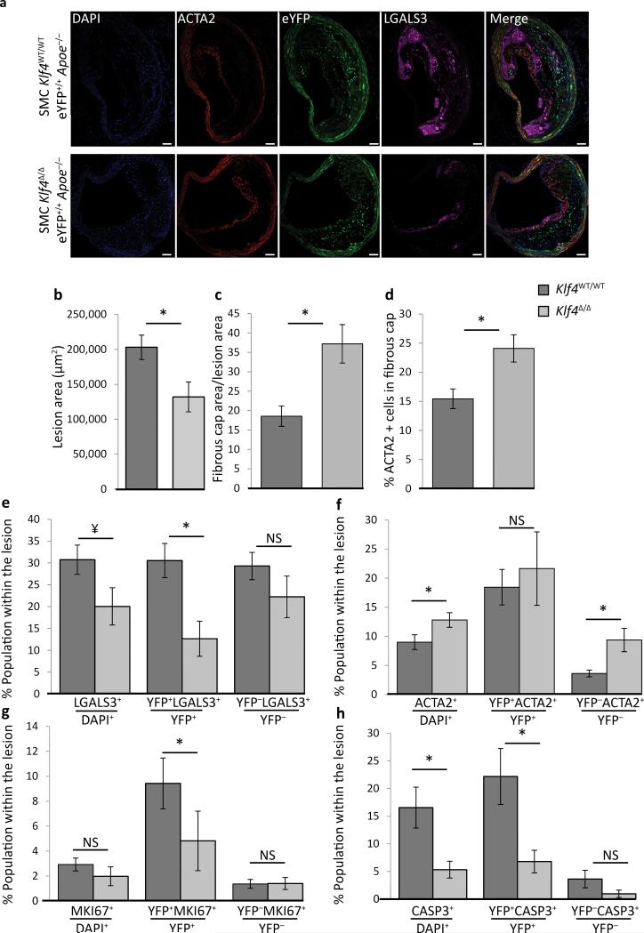
Figure 4. SMC specific Klf4 conditional KO in Apoe−/− mice fed a high fat diet for 18 weeks resulted in decreased lesion size and increased indices of plaque stability.
SMC eYFP+/+ Apoe−/− mice crossed to Klf4FL/FL mice were tamoxifen treated and given Western diet for 18 weeks prior to analysis. (a) Representative image demonstrating the changes in lesion morphology and cell composition. Results showed the following: (b) decreased total lesion area, (c) increased fibrous cap area (defined as the region of the lesion within 30 μm of the luminal surface) relative to the size of the lesion, (d) increased ACTA2+ cells within the fibrous cap, (e) an overall decrease in the fraction of LGALS3+ cells, and a large decrease in the fraction of SMC-derived LGALS3+ cells (YFP+LGALS3+), (f) an increase in the ACTA2+ population within the lesion, (g) decreased proliferation of SMC-derived cells (YFP+MKI67+), and (h) decreased overall cell death, mostly due to a decrease in SMC-derived apoptosis (YFP+CASP3+). *P < 0.05, ¥ P = 0.07, analysis completed by 2-way ANOVA comparing genotyping and distance from start of the BCA with a Tukey post-test, error bars are based on S.E.M. Quantification is based on analysis of five 71415 μm2 regions in each of three sections (d-g), or two sections (h, i), per mouse spanning a 600μm length of the BCA. Scale bar = 50 μm. SMC YFP+/+ Klf4WT/WTApoe−/− n = 11, SMC YFP+/+ Klf4Δ/ΔApoe−/− n = 8.

