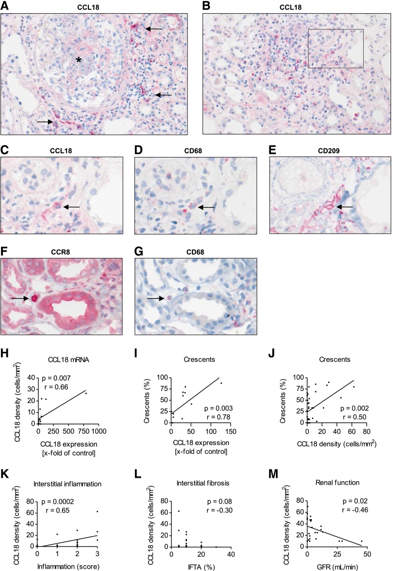
Figure 3.
Immunohistochemistry reveals the presence of mononuclear intrarenal CCL18+ cells, which correlate with renal function at the time of biopsy. (A and B) Representative CCL18 immunohistochemistry of a renal biopsy from a patient with ANCA-associated crescentic GN showed that CCL18+ cells were predominantly located in the periglomerular and inflamed, nonscarred tubulointerstitium. Two-micrometer-thick FFPE tissue sections were stained for CCL18. Arrows indicate the CCL18+ cells surrounding a glomerulus marked with an asterisk (magnification, ×200). (C) CCL18, (D) CD68, and (E) CD209 staining revealed the presence of CCL18+ cells coexpressing CD68 and CD209. Arrows indicate a CCL18+ CD68+ CD209+ myeloid DC on the basis of staining of consecutive sections with CD68 and CD209 (original magnification, ×400). (F) Two-micrometer-thick FFPE tissue sections of patients with renal ANCA disease were stained for CCR8, identifying mononuclear CCR8+ cells with the appearance of macrophages. (G) Staining of consecutive sections with (F) CCR8 and (G) CD68 revealed the presence of CCR8+ cells coexpressing CD68 (original magnification, ×400). Arrows indicate a CCR8+ CD68+ macrophage on the basis of staining of consecutive sections with CD68. (H) The relative expression of CCL18 mRNA correlated with the density of CCL18+ cells (n=15). (I) The CCL18 mRNA levels correlated with the percentage of cellular crescents (n=12). (J) The percentage of cellular crescents correlated with the number of CCL18+ cells (n=31). (K) The density of CCL18+ cells correlated with the degree of acute interstitial inflammation (n=31). (L) The number of CCL18+ cells did not correlate with interstitial fibrosis and tubular atrophy (IFTA; n=31). (M) The density of the CCL18+ cells correlated with impairment of renal function (GFR) in patients who did not receive immunosuppressive treatment before renal biopsy (n=24).

