Figure 1.
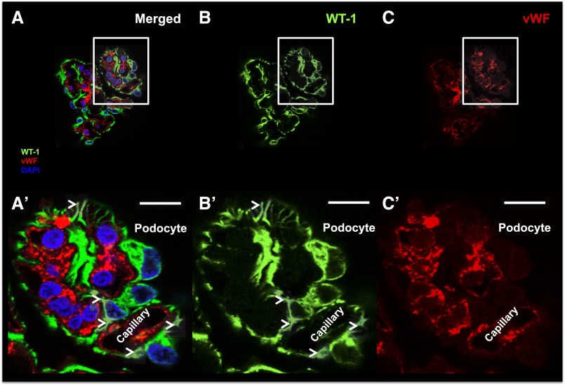
Podocyte identification. (A) A representative confocal image of a glomerular tuft is shown (merged) using (B) WT-1 (green; specific podocyte marker), (C) vWF (red; specific endothelial cell marker), and DAPI (blue; nuclear marker). The corresponding insets show our podocyte identification criteria: (A′) expression of WT-1 in podocyte cytoplasm, (B′) lack of expression of vWF, and (C′) their location outside capillaries. Arrowheads show classic podocyte morphology (major projections). Scale bars, 10 μm. DAPI, 4′,6-diamidino-2-phenylindole.

