
Figure 2.
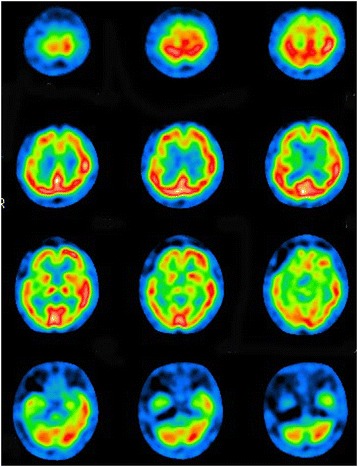
Single photon emission computed tomography imaging revealed obvious hypoperfusion of the right frontal and temporal cortices, basal ganglia and brainstem, while perfusion of right cerebellar cortex was mildly impaired.
Official websites use .gov
A
.gov website belongs to an official
government organization in the United States.
Secure .gov websites use HTTPS
A lock (
) or https:// means you've safely
connected to the .gov website. Share sensitive
information only on official, secure websites.
Single photon emission computed tomography imaging revealed obvious hypoperfusion of the right frontal and temporal cortices, basal ganglia and brainstem, while perfusion of right cerebellar cortex was mildly impaired.