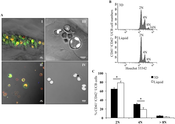
Fig 6. In situ characterization of differentiation markers and ploidy of mature MK.
(A) Immunofluorescence staining of CD41 (green) and CD42b (red) cells growing inside pores of 3D (I) and in liquid culture (II). Nuclear staining of cell grown in 3D (III) and liquid culture (IV) with YOYO-1 marker (white). All images were acquired 12 days after seeding using the Leica 510 confocal microscope with 40X Plan-NeoFluar objective lens. Scale bar = 10 μm. (B) Representative flow cytometry ploidy analysis of CD41+/CD42b+ UCB cells from 3D and liquid culture, 11 days after seeding. (C) Ploidy analysis of CD41+/CD42b+ UCB cells in 3D (black bars) compared to liquid culture (white bars), 11 days after seeding. Data are means ± SEM of 3 independent experiments. *p<0.05. Abbreviation: UCB, umbilical cord blood.

