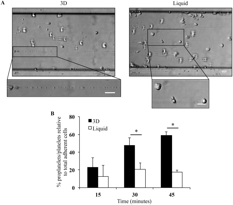
Fig 7. MK deformation and platelet production in flow conditions.
(A) Stages of MK deformations and reorganization into proplatelets and platelets at 20 minutes of perfusion in 3D (left panel) and in liquid culture (right panel) using Bioflux microfluidic platform to increase platelet formation from mature MK [10]. Images were acquired using the Axiovert 135 transmission optical microscope with 20X Plasdic magnification. Bar = 20 μm. (B) Histogram representation of proplatelet/platelet numbers as a percentage of total adherent cells per field recovered from mature MK obtained in 3D (black bars) and liquid culture (white bars). Data are means ± SEM of 3 independent experiments.*p<0.05.

