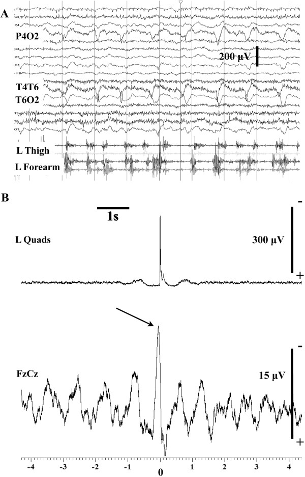
Figure 1.

Myoclonic status epilepticus (case 7). (A) Routine EEG while the patient was obtunded and had frequent irregular left upper and lower limb myoclonic twitches. The surface EMG electrodes in the left thigh were recording from the quads (upper trace) and the biceps femoris. In the forearm, surface electrodes were recording from the extensor digitorum communis (upper trace) and the finger flexors. EEG tracing shows, about 1 Hz, periodic lateralized epileptiform discharges in the right posterior quadrant. Note that these are not time matched to the myoclonic jerks from the left arm and leg. (B) Jerk-locked back averaging from the left quads (80 sweeps). There is a time locked negative sharp wave preceding the onset of the averaged, rectified EMG data by about 25 ms. These recordings were performed in the ICU at a sampling rate of 500 Hz.
