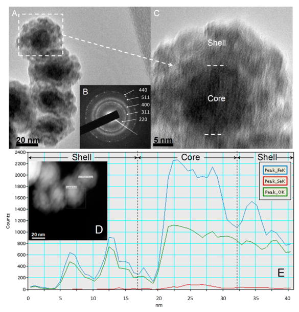
Fig. 11.
TEM images of iron nanoparticles after selenate removal in DI water (five cycle reaction). (A) Bright field image; (B) Selected area electron diffraction (SAED); (C) High-resolution TEM of iron/iron oxide core-shell structure; (D) Dark field image in STEM mode; (E) STEM-EDS line scan spectrum (step size: 1.5 nm, pixel time: 30s). For each cycle, the reaction time was 10 min. [Fe]0=0.50±0.02 g/L, [Se]0=2.0±0.2 mg/L (made by Na2SeO4), and pH was adjusted to 4.5. The dash lines separate the core and shell of particles. Blue/red/green lines indicate Fe/Se/O, respectively.

