
Figure 1.

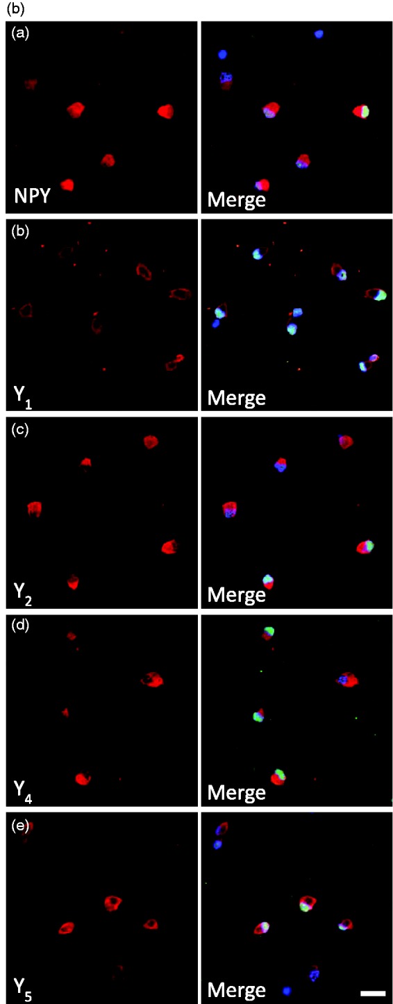

Expression of NPY and NPY receptors in RGCs. (a) Detection of NPY and NPY receptors (Y1, Y2, Y4, and Y5) mRNA expression by RT-PCR in purified RGCs. RGCs were isolated from the retinas of either pups or young adult Long Evans and Wistar rats. DNA ladder. (b) NPY and NPY receptor immunoreactivity (ir) in a purified RGC culture obtained from Wistar pups (a–e, red). RGCs were stained with antiBrn3a (RGC marker, green) and nuclei with DAPI (blue). Scale bar: 20 µm. (c) NPY-ir and NPY receptor-ir in retinal slices obtained from young adult Wistar rats (a–e, red). RGCs were stained with the RGC marker Brn3a (green) and nuclei with DAPI (blue). NPY-ir was mainly detected in strata 1, 3, and 5 of IPL (arrows; a). Y1-ir was detected in distal and proximal INL, strata 2 and 4 of IPL, and RGCs (arrows; b). Y2-ir was detected in INL (c). Y4-ir was detected in INL and GCL (d). and Y5-ir was detected in Müller cells (e). (d) Y5-ir (red) is colocalized with Müller cells (green) in retinal slices of young adult Wistar rats. Müller cells were identified by vimentin-ir (green). Nuclei were stained with DAPI (blue). GCL = ganglion cell layer; IPL = inner plexiform layer; INL = inner nuclear layer; OPL = outer plexiform layer; ONL = outer nuclear layer. Scale bar: 50 µm.