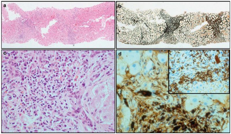
Figure 2.
Fibro-inflammatory changes in the liver. Liver biopsy (a: hematoxylin and eosin (H&E), ×40 and b: reticulin, ×40) showing thick fibrous bands with nodule formation. Inflammatory cell infiltrate rich in lymphocytes and plasma cells (c: H&E ×400). Large number of plasma cells expressing IgG4 (d: IgG4, ×400; inset: CD138, ×200).

