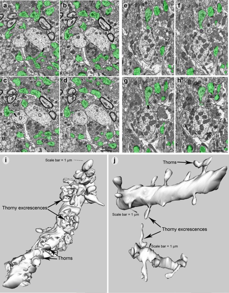
Figure 3. 3-D reconstruction of CA3 pyramidal cell dendrites reveals loss of thorny excrescences in Tc1 mice.
Consecutive images of proximal portions of CA3 dendrites for both wild-type (a-d) and Tc1 mice (e-h). Thorny excrescence profiles are labelled in green, exemplifying a clear reduction of thorns in Tc1 mice. Abbreviations: D, dendritic shafts; GB, mossy fiber giant boutons or varicosities representing pre-synaptic portions of the synapses; MF, mossy fibers or axons originating from granule cells. 3-D reconstructed CA3 dendritic segments with thorny excrescences in (i) wild-type and (j) Tc1 mice.

