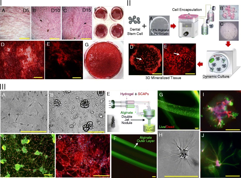
Figure 3.

Osteogenic and neural differentiation of mycoplasma-eliminated hSCAPs. I. 2D Osteogenic differentiation of mycoplasma-eliminated hSCAPs: A, B and C. Phenotypic ALPase expression after 5, 10 and 15 days of osteogenic culture, D, F and G. Alizarin red-S stained image of mineralized nodules after 20 days of osteogenic culture under light microscope, E. alizarin red-S stained image of mineralized nodules after 20 days of osteogenic culture under fluorescence microscope, II. 3D Osteogenic differentiation of mycoplasma-eliminated hSCAPs: A. alginate hydrogel, B and C: alginate hydrogel encapsulating hSCAPs, D and E. alizarin red-S stained image of mineralized nodules within alginate hydrogel after 20 days of 3D osteogenic culture under fluorescence microscope, III. 2D and 3D neural differentiation of mycoplasma-eliminated hSCAPs: A and B. microscopic images of hSCAPs in 2D neural differentiation culture. C and D. βIII tubulin (red), Cam kinase II (green) and nuclear staining (DAPI; blue), E. Schematic illustration of hSCAPs encapsulation of hSCAPs for 3D neural differentiation. F. tubular alginate hydrogel, G. live and dead image of encapsulated hSCAPs in tubular alginate hydrogel, H. microscopic images of hSCAPs in 3D neural differentiation culture, I. βIII tubulin (red), Cam kinase II (green) and nuclear staining (DAPI; blue) of differentiated hSCAPs in 3D neural differentiation culture, J. βIII tubulin (green), Cam kinase II (red) and nuclear staining (DAPI; blue) of differentiated hSCAPs in 3D neural differentiation culture, Scale bars indicate 200 μm.
