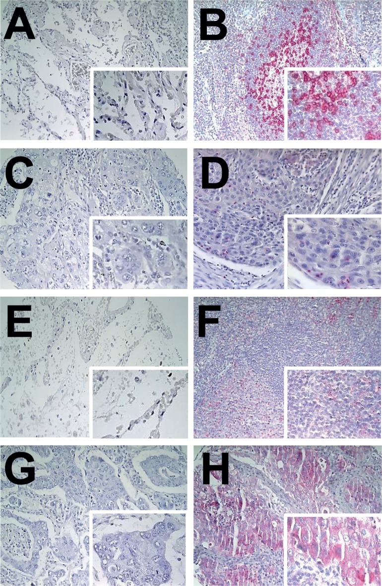
Fig 1. Representative immunohistochemical staining results for PD-1 (A: normal lung tissue, negative control; B: tonsillar tissue, positive control; C: PD-1-negative tumor infiltrating lymphocytes; D: PD-1-positive tumor infiltrating lymphocytes in squamous cell carcinomas) and for PD-L1 (E: normal lung tissue, negative control; F: tonsillar tissue, positive control; G: PD-L1 negative squamous cell carcinomas; H: PD-L1 positive squamous cell carcinomas).

All images at x20, inlay x40.
