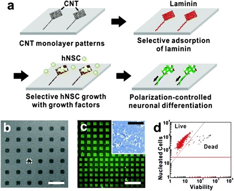
Figure 2.

Shape-controlled CNT substrates for hNSCs growth and polarization. (a) Schematic diagram showing the process of the polarization-controlled neuronal differentiation of hNSCs. Shape-controlled CNT substrates induced the differentiation of hNSCs into neuronal lineages. (b) SEM image of CNTs substrate. Scale bar represents 40 μm. (c) Immunofluorescence image of anti-laminin (green) bound to the laminin absorbed on the CNT substrate. Scale bar represents 200 μm. The inset shows AFM image of laminin-coated CNT substrate. Scale bar of the inset represents 2 μm. (d) Cell viability of hNSCs cultured on CNT substrates for 3 day proliferation. The data indicates that 98% of hNSCs cultured on CNT substrates were alive (red) (adapted from reference [21]).
