Figure 3.
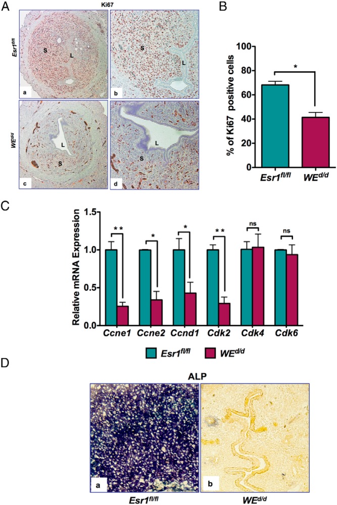
Uterine stromal cell proliferation during decidualization is impaired in WEd/d mice. A, Cell proliferation was measured by Ki67 immunostaining in uterine sections of Esr1fl/fl (upper panel) and WEd/d (lower panel) mice at 20 hours after decidual stimulation. Magnification (a and c), ×10; (b and d), ×20. B, Percentage of Ki67-positive cells in the stromal compartment of Esr1fl/fl and WEd/d mice 20 hours after decidual stimulation. The data represent the average number of cells from multiple fields of multiple uterine sections. *, P ≤ .05 (t test, n = 4). C, Stromal cells were isolated from uteri 20 hours after decidual stimulation and total RNA was prepared. Real-time RT-PCR was performed to monitor the expression of mRNAs corresponding to the cell cycle genes Ccne1, Ccne2, Ccnd1, Cdk2, Cdk4, and Cdk6. Rplp0 encoding a ribosomal subunit protein was used as internal control to normalize gene expression. The data are represented as the mean fold induction ± SEM. *, P ≤ .05; **, P ≤ .01. ns, not significant. D, ALP activity was detected in the decidualized stroma. a, sections of Esr1fl/fl uteri; b), sections of WEd/d uteri. Purple color indicates ALP activity.

