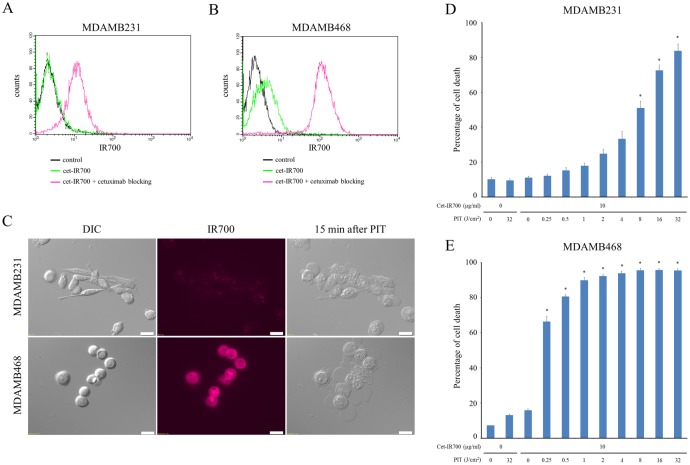
Fig 1. Confirmation of EGFR expression as a target for PIT in MDAMB231 and MDAMB468 cells, and evaluation of in vitro PIT.
(A) Expression of EGFR in MDAMB231 cells was examined with FACS. (B) Expression of EGFR in MDAMB468 cells was examined with FACS. (C) MDAMB231 and MDAMB468 cells were incubated with cet-IR700 for 6 h and observed by microscopy. Fluorescence intensities of MDAMB468 were higher than MDAMB231. Necrotic cell death was observed upon excitation with 2 J/cm2 of NIR light (after 15min). Bar = 20 μm. (D) Membrane damage of MDAMB231 cells induced by PIT was measured with PI staining (dead cell count), which increased in a light dose-dependent manner (n = 5, *p < 0.001, vs. untreated control, by Student’s t test). (E) PI staining showed membrane damage of MDAMB468 cells (n = 5, *p < 0.001, vs. untreated control, by Student’s t test).

