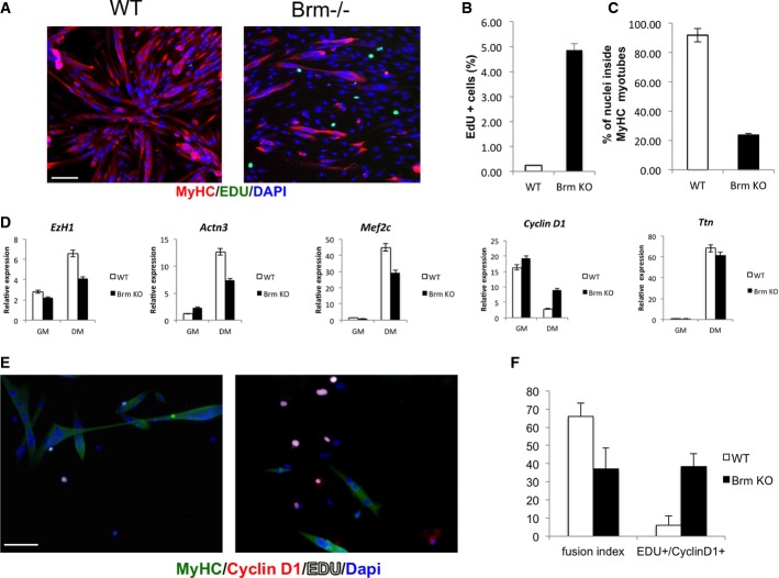
Proliferation and differentiation defect in Brm−/− satellite cells
Satellite cells were isolated from muscles of WT and Brm−/− mice, cultured in GM, and then exposed to DM. Cells were incubated with EdU for 6 h in DM prior to collection for staining.
- A Immunofluorescence staining of MyHC and EdU. Scale bar, 50 μm.
- B Percentage of EdU-positive cells was calculated counting 10 fields of EdU-positive cells.
- C Quantification of fusion index calculated as percentage of nuclei within MyHC-expressing myotubes.
- D Analysis of expression levels of transcripts of genes selected from microarray analysis.
- E, F Immunofluorescence staining of WT (left)- and Brm−/− (right)-derived satellite cells for MyHC, EDU, and cyclin D1 (E); relative quantification of fusion index; and number of EDU+/cyclin D1+ cells (F). Scale bar, 50 μm. Data are shown as average ± SEM (n = 3).

