Figure 2.

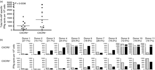
(a) Quantification of influenza-reactive circulating follicular helper T (Tfh) and non-Tfh cells. CXCR5+ and CXCR5– CD4+ CD45RA− T cells were cultured with 2 μm pools of peptide for haemagglutinin (HA) or nucleoprotein (NP) influenza proteins in the presence of autologous antigen-presenting cells (APC). Antigen-specific CD4 T cells were measured by interferon-γ (IFN-γ) EliSpot at 36–40 hr of stimulation and the abundance was summed. (b) Influenza protein-specific reactivity in circulating Tfh and non-Tfh. Frequency of HA-specific (grey bars) or NP-specific (black bars) IFN-γ secreting cells per 1 000 000 sorted CXCR5– (top row) or CXCR5+ (bottom row) for individual donors. The percentage of CXCR5+ circulating Tfh found in each subject is indicated below the subject number at the top of each panel.
