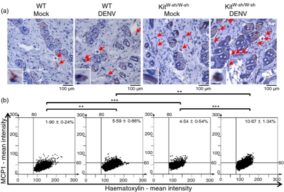
Figure 5.

Expression levels of CCL2 (MCP-1) at the skin inoculation site are higher in KitW-sh/W-sh than in wild-type (WT) mice. WT and KitW-sh/W-sh mice (n = 8/group) were intradermally (i.d.) inoculated with medium (Mock) or dengue virus (DENV) (1 × 10⁹ plaque-forming units/mouse) at four sites on the upper back and were killed 3 days post-infection (d.p.i.). (a) The skin inoculation site sections were stained with anti-CCL2 (MCP-1) antibody (red) and nuclei were stained with haematoxylin (blue). Red arrows indicate CCL2 (MCP-1)-positive cells (magnification: ×200). (b) The CCL2 (MCP-1)-positive cells were counted in 15 regions per mouse field and the average numbers of CCL2 (MCP-1)-positive cells were calculated using HistoQuest software. **P < 0·01; ***P < 0·001.
