Figure 4.

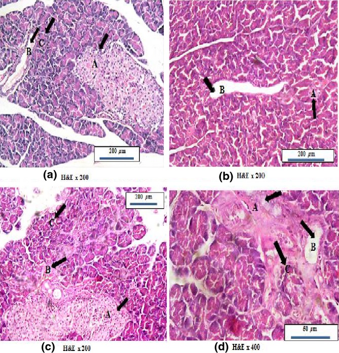
Photomicrographs of rat pancreas stained with H&E; (a) Control pancreas showing normal acinar arrangement with basal basophilia and apical acidophlia (black arrow &C) and normal sized interlobular duct (black arrow &B), with normal sized islet of Langerhans (black arrow &A). (b) Pancreas of high salt diet fed group showing widened interlobular duct (black arrow &B) and degenerated islet of Langerhans (black arrow &A). (c) Pancreas of high salt diet fed group showing degenerated acini (black arrow &C) with burden interlobular duct (black arrow &B) inside C.T septa in addition to fused islets of Langerhans (black arrow &A). (d) Pancreas of high salt diet fed group showing expanded fibrous tissue septa with disruption of acinar arrangement appeared as entrapped acini (black arrow &C) with widened interlobular duct (black arrow &B) and also degenerated entrapped islet of Langerhans (black arrow &A).
