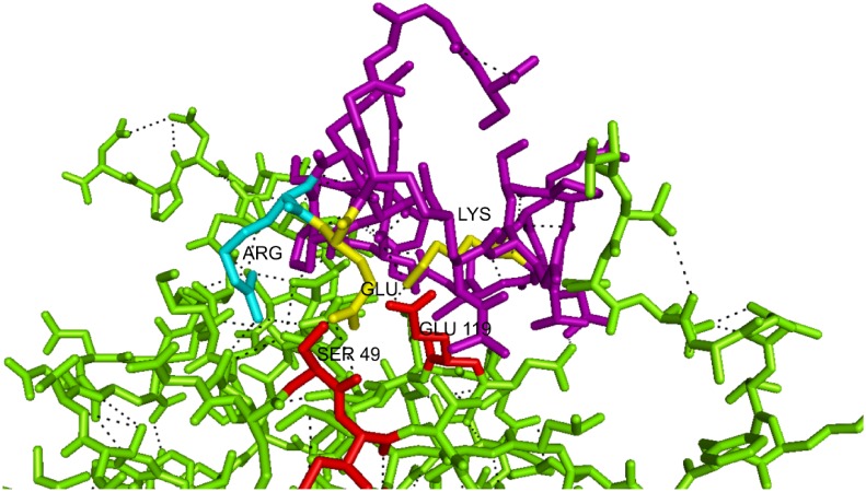
Fig 3. Three-dimensional analysis showing the region of interaction between the pm26TGF-β1 peptide and TβRII.
The interaction between the Glu and Lys residues present in the pm26TGF-β1 peptide (purple) and the Ser49 Glu119 residues of the TβRII (red). The binding site shared by TGF-β1 and pm26TGF-β1 peptide is represented in yellow. The Arg residue present in the pm26TGF-β1 peptide is represented in cyan.

