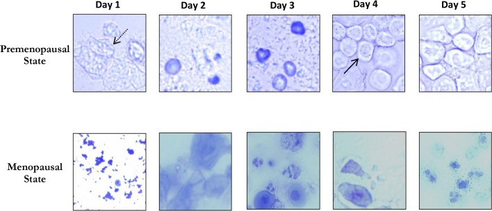
Fig 4. Representative examples of vaginal smears obtained from premenopausal (the upper images) and menopausal (the bottom images) female rats aged 18 months collected over five consecutive days.
A typical estrus cycle (with the four consecutive phases: proestrus, estrus, metestrus, and diestrus) could be identified in premenopausal rats. Proestrus phase was identified with the presence of nucleated epithelial cells (solid line in image taken at day 4); Estrous phase was recognized with the presence of cornified epithelial cells (dashed line in image taken at day 1); the other two phases (Metestrus and Diestrus) were mainly distinguished with the leukocyte infiltration (images taken at day 2 and 3). No equivalent distinct estrus phase could be observed in menopausal group. The images were taken with a 40X objective.

