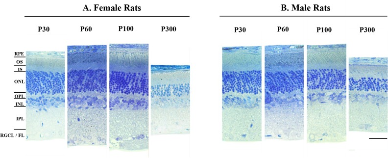
Fig 6. Representative retinal sections obtained at 30, 60, 100, and 300 days of age from female (A) and male adult SD rats (B).
Images were taken between 1020 μm and 1700 μm from the optic nerve head in the superior retina. Abbreviations: RPE: retinal pigmented epithelium; OS: outer segment; IS: inner segment; ONL: outer nuclear layer; OPL: outer plexiform layer; INL: inner nuclear layer; IPL: inner plexiform layer; RGCL/FL: retinal ganglion cell layer/fiber layer. Calibration bar: 75μm.

