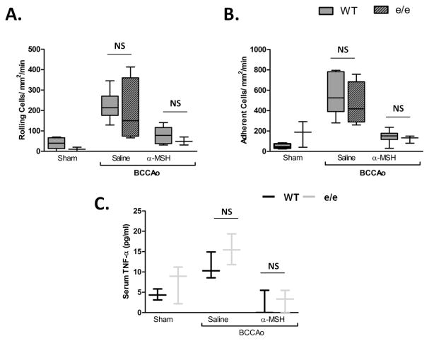
Figure 5. The e/e mouse inflammatory phenotype is diminished by 2h.
WT (C57BL/6) (white bars) and e/e (lined bars) mice were subjected to sham or BCCAo and treated with saline or 10μg α-MSH at the start of reperfusion. Leukocyte recruitment was quantified at 2h of reperfusion in terms of: in terms of: A) rolling cell flux and B) leukocyte adhesion (cells/mm2/min). n = 4 mice/group for e/e saline group, and n=3 mice/group for sham and α-MSH e/e groups, comparisons were made to WT groups as in figure 1 (n=6 mice/group). Statistical analysis was performed between mouse strains, within the same treatment group to evaluate phenotypic differences between strains. NS denotes no statistical significance to WT group. C) ELISA measurements of serum TNF-α in WT and e/e mice. * denotes significance to sham groups P<0.01, † denotes significance to BCCAo P<0.05.

