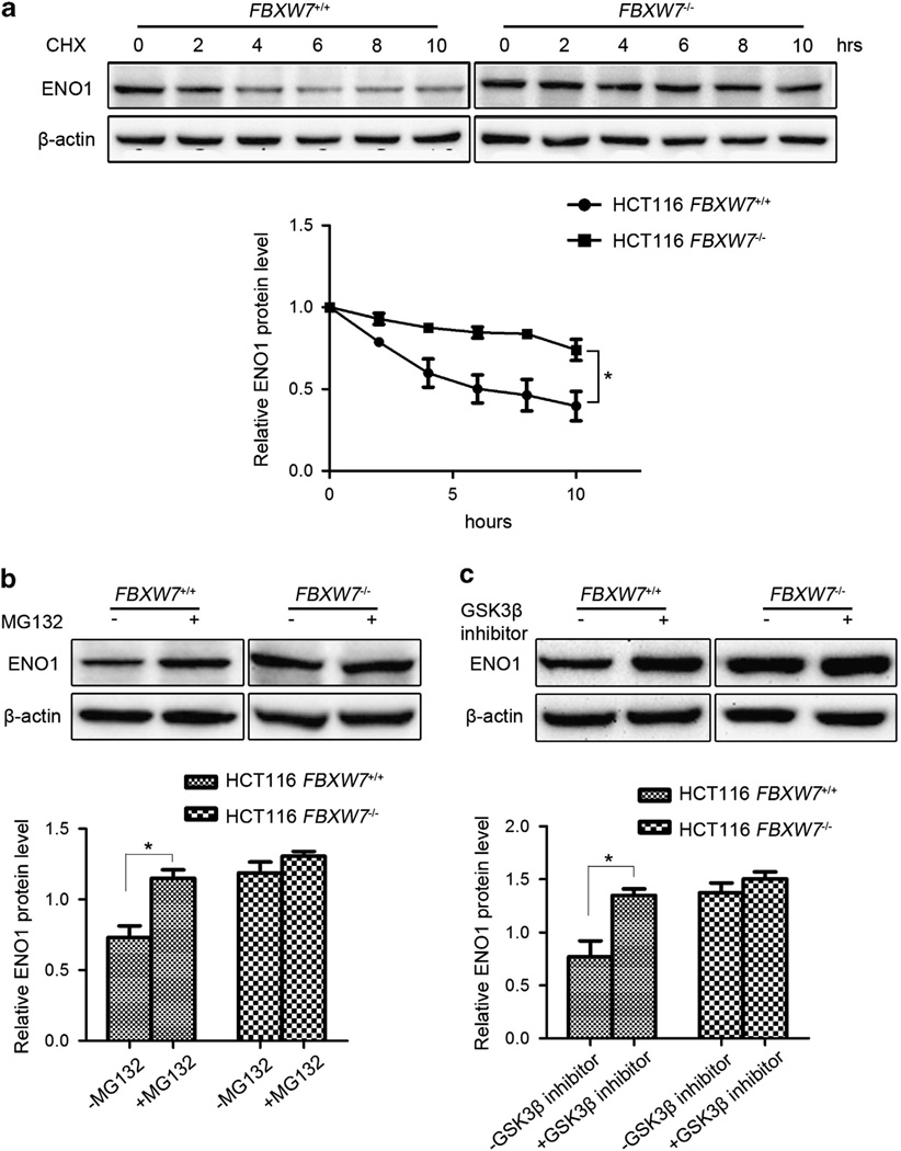
Figure 3.
FBXW7 facilitates ENO1 turnover in proteasome-dependent pathway. (a) HCT116 FBXW7+/+ and FBXW7−/− cells were treated with 50 µg/ml CHX for 0, 2, 4, 6, 8 and 10 h. Western blotting analysis was carried out to detect the endogenous level of ENO1 using the anti-ENO1 antibody. The graph shows quantitative analysis of the CHX chase data. (b) HCT116 FBXW7+/+ and FBXW7−/− cells were treated with or without 10 µM MG132 for 6 h. Western blotting analysis was carried out using the anti-ENO1 antibody. The graph shows quantitative analysis. (c) HCT116 FBXW7+/+ and FBXW7−/− cells were treated with or without 25 µM GSK3β inhibitor VIII for 6 h. Western blotting analysis was carried out using the anti-ENO1 antibody. The graph shows quantitative analysis. All results are representative of three independent experiments. *P < 0.05 based on Student’s t-test.

