Figure 3.
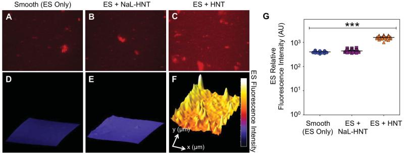
Detection of immobilized ES on biomaterial surfaces. (A-C) Representative high magnification fluorescence micrographs of recombinant human ES (red) adsorbed on smooth (ES only; A) surfaces, immobilized NaL-HNT (B), and HNT (C) coated microscale flow devices. Scale bar = 40 μm. (D-F) Representative three-dimensional surface plots of immobilized recombinant human ES fluorescence intensity on smooth (D), NaL-HNT (E), and HNT (F) coated microscale flow devices. Profile length in x- and y-directions are 240 μm and 160 μm, respectively. (G) Immobilized ES relative fluorescence intensity values on smooth and nanostructured surfaces. Calculated values are mean +/- standard deviation (n = 3). Statistics were calculated using a one-way ANOVA with Tukey post test. ***P < 0.0001.

