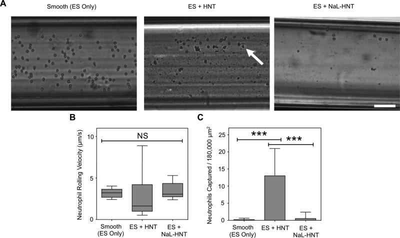
Figure 5.
ES-mediated adhesion of leukocytes to immobilized surfactant-nanotube complexes under flow. (A) Representative images of ES-mediated adhesion of primary human neutrophils under flow on smooth surfaces, immobilized HNT, and NaL-HNT coated microscale flow device biomaterial surfaces. Arrows denote adhered neutrophils, which exhibit either rolling or firm adhesion. Scale bar = 100 μm. (B) Neutrophil rolling velocities on ES on smooth, HNT, and NaL-HNT coated microscale flow device surfaces. Error bars denote minimum and maximum data points. Statistics were calculated using a two-tailed unpaired t-test. NS: not significant. n = 30 or more rolling cells analyzed for each condition. (C) Number of captured neutrophils per 180,000 μm2 of biomaterial surface area. Captured neutrophils denote cells that are firmly adhered to the surface. Calculated values are mean +/- standard deviation. n = 20 or more frames analyzed for captured cells for each condition. Statistics were calculated using a one-way ANOVA with Tukey post test. ***P < 0.0001.

