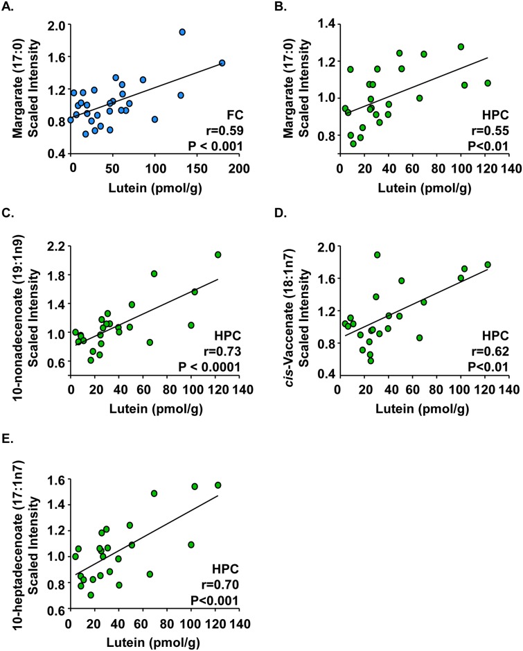
Fig 2. Fatty acids correlated with lutein concentrations in infant brain.
Post-mortem infant brain tissues (age 1 to 488 days) from the frontal cortex, hippocampus, and occipital cortex were analyzed for both lutein and lipid pathway metabolites. Results are shown as metabolite (scaled intensity) by lutein concentration (pmol/g). In the frontal cortex (n = 29), (A) margarate had a strong, positive correlation with lutein. In the hippocampus (n = 24), (B) margarate, (C) 10-nonadecenoate, (D) cis-vaccenate, and (E) 10-heptadecenoate all had strong, positive correlations with lutein. No strong correlations were observed in the occipital cortex. Lipid pathway metabolite:lutein correlations with r values ≥ |0.6| and P < 0.05 are shown. FC: frontal cortex; HPC: hippocampus.

