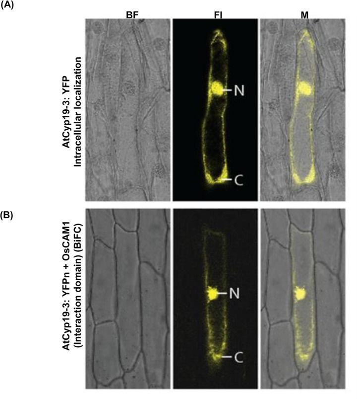
Fig 8. Intracellular localization of AtCyp19-3.
The upper panel (A) shows the accumulation pattern of AtCyp19-3:YFP chimeric protein. The lower panel (B) shows the sub-cellular interaction of AtCyp19-3-1:YFPn and OsCaM1:YFPc chimeric proteins by bi-molecular fluorescence complementation. The chimeric polypeptide containing AtCyp19-3(71–176), (which did not show binding to CaM by gel overlay assay) with YFPn served as a negative control for in vivo studies. BF: bright field; Fl: fluorescence image; M: merged image, N: nucleus; C:cytoplasm.

