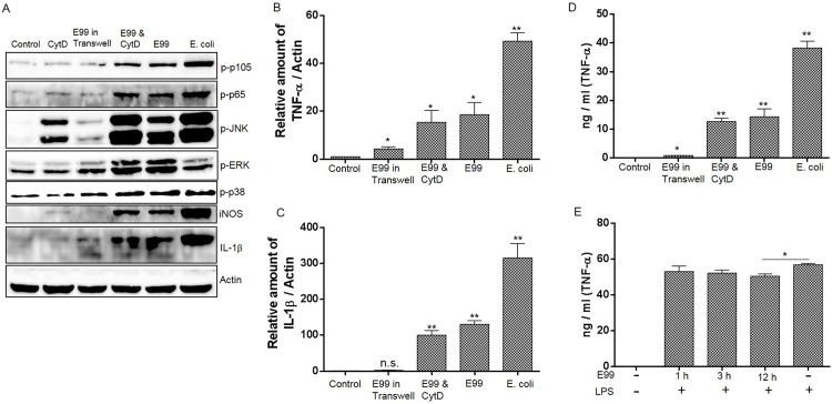
Fig 3. The effect of E. faecalis on NF-κB and MAPK activation and cytokine expression.
This experiment included the following groups: (1) RAW264.7 cells alone; (2) RAW264.7 cells infected with E. faecalis E99; (3) RAW264.7 cells infected with E. coli; (4) E. faecalis E99 were added on the top of a Transwell semipermeable filter separating the bacteria from RAW264.7 cells; (5) the RAW264.7 cells were pretreated with CytD for 0.5 h before being infected with E. faecalis E99 for 5 h. The cells were collected to analyze the activation of NF-κB and MAPK by Western blot (A), or to measure the mRNA level of TNF-α (B) and IL-1β (C) by RT-PCR. (D) The supernatant from treated cells above were used to measure the concentration of TNF-α by ELISA. *, p<0.05; **, p<0.01 represent statistically significant difference compared to RAW264.7 cells without infection; n.s., not statistically significant compared to RAW264.7 cells without infection. (E) RAW264.7 cells were infected with E99 for 1h, cells were washed thrice with PBS and further incubated with medium containing vancomycin and gentamicin to kill the extracellular bacteria for 1 h, 3 h or 12 h before stimulation with LPS (1μg/ml) for 3.5 h. The supernatants were collected for analysis of TNF-α concentration by ELISA. *, p<0.05.

