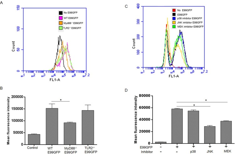
Fig 6. Impaired phagocytosis in the absence of MyD88, ERK and JNK signal pathway.
(A) WT, TLR2-/-, MyD88-/- BMDM were infected with E99GFP at a MOI of 100 for 1 h, then the cells were analyzed by FACS after washing thrice with PBS. Representative FACS histogram shows the phagocytosis of E99GFP by WT, TLR2-/- and MyD88-/- BMDM. (B) Mean fluorescence intensity of GFP from A. *, p<0.05. (C) RAW264.7 cells were pretreated with inhibitors of p38, JNK or MEK and then infected with E99GFP at MOI of 100 for 1 h. The cells were washed with PBS for three times before analysis by FACS. FACS histogram shows phagocytosis of E99GFP by RAW264.7 cells with different treatments. (D) Mean fluorescence intensity at 1 hour after phagocytosis of E99GFP by RAW264.7 cells from C. *, p<0.05;

