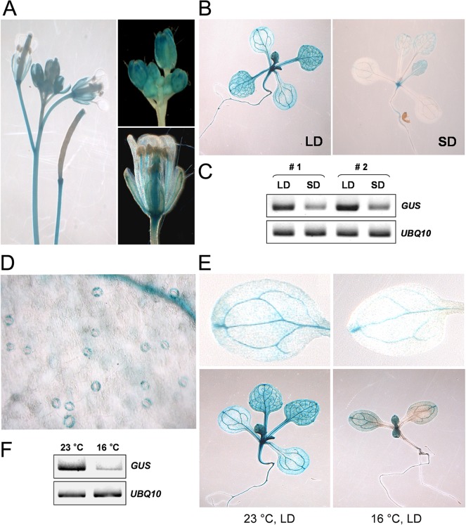
Fig 6. A homozygous line expressing a PaFT1-promoter GUS fusion that showed a characteristic staining pattern was chosen for histochemical analysis of pPaFT1:GUS expression in different organs of Arabidopsis.
A, Inflorescence, floral buds, flower and siliques. B, 8-day-old seedlings grown under LD and 14-day-old seedlings grown under SD. C, GUS mRNA expression under different photoperiods. D, GUS expression in guard cells. E and F, Different levels of GUS expression are detectable under different temperature. 8-day-old seedlings grown under constant 23°C and 16°C, respectively were used for GUS staining or mRNA analysis.

