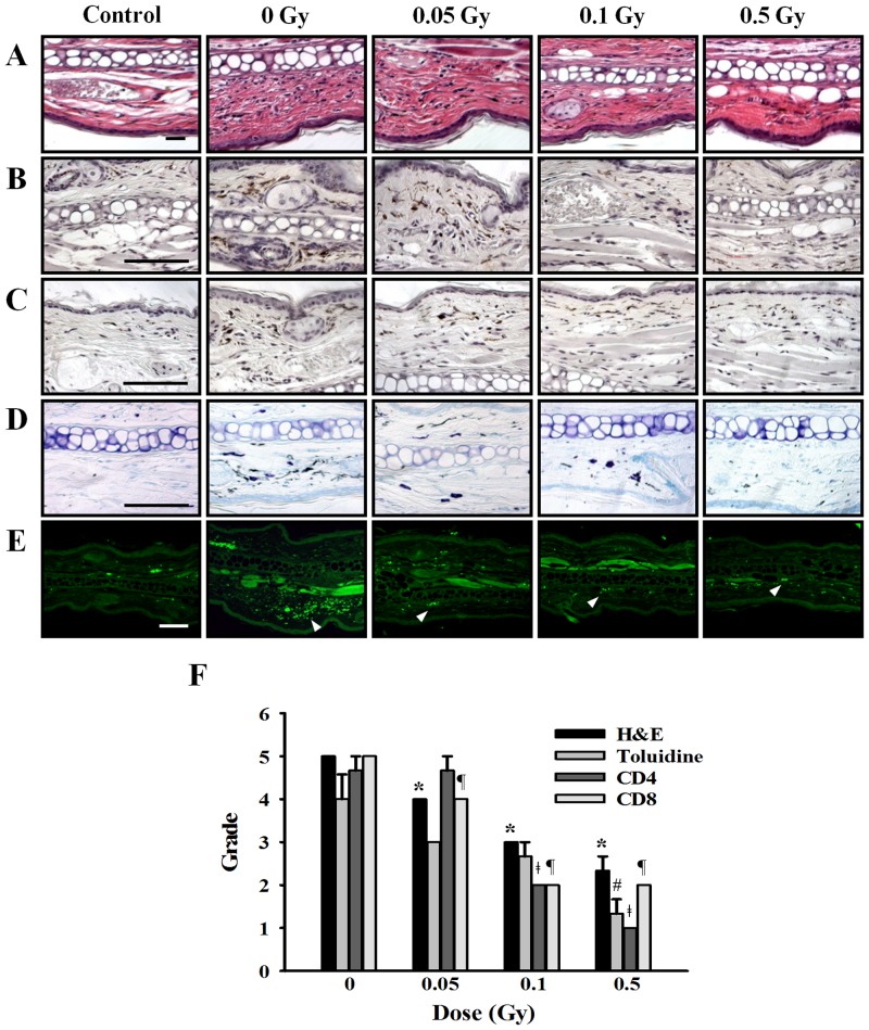
Fig 8. Effects of ionizing radiation on histological changes in ear tissue of passive cutaneous anaphylaxis model.
Anti-DNP (20 ng) was injected intradermally in the left ear, whereas the right ear was injected with saline as a control. After 24 h, mice were irradiated with 0–0.5 Gy before injection of 100 μg DNP-HAS via the tail vein. After 5 hr, sections of ear tissue were stained with H&E (A), immunohistochemistry for CD4 (B) and CD8 (C), toluidine blue (D), and FcεRI expression (E) observed at 400× magnification. Histological appearance was score for the presence of infiltrating cells (F). Bars = 100 μm. * p < 0.05, # p < 0.05, ǂ p < 0.05, ¶ p < 0.05 versus the corresponding 0 Gy of H&E, Toluidine, CD4, and CD8, respectively.

