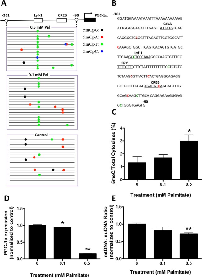
Fig 5. Palmitate induces PGC−1α promoter methylation in primary astrocytes.

Primary astrocytes were treated with 0.1 or 0.5 mM Palmitate (Pal) for 48 hours. Genomic DNA and mRNA were isolated for bisulfite sequencing and RT-PCR analysis. A. Graphical depiction of the PGC−1α promoter region (-361 to -90) and location of methylated cytosines. B. Methylation sequencing region in the PGC−1α promoter. Important transcription factors are underlined. Methylated CpGs are highlighted in brown, CpAs in red, CpTs in green and CpCs in blue. C. Quantitation of cytosine methylation levels of the PGC−1α promoter. Methylation of the PGC−1α promoter was increased by 129.2% with the treatment of 0.5 mM palmitate (p = 0.012) compared to PBS control. D. Quantification of PGC−1α mRNA by qRT-PCR revealed a 6.4% decrease with the treatment of 0.1 mM palmitate (p = 0.0085) and 83.9% decrease with the treatment of 0.5 mM palmitate (p<0.0001) compared with PBS controls. E. Quantitation of the mitochondria DNA (mtDNA) to nuclear DNA (nuDNA) ratio using real-time PCR. mtDNA:nuDNA ratio was decreased by 28.1% in 0.5 mM palmitate-treated astrocyte cultures compared to control (p = 0.0006). Results presented as mean±SEM, *p<0.05, **<0.01, ANOVA with Student-Newman-Keuls post hoc analysis.
