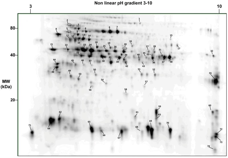
Fig 3. 2D-gel reference map of the short ragweed pollen proteome.
Proteins from an aqueous short ragweed pollen extract were separated by 2D-gel electrophoresis and stained with Sypro Ruby. Proteins spots were picked and analyzed by LC-MS/MS after trypsin digestion. Proteins were identified using the Transcriptome-Derived Proteome collection supplemented with missing known allergens. Numbers refer to spots analyzed by mass spectrometry. Identification details are provided in Table 1.

